ACCS-EM · Emergency Medicine
Surviving to Thriving
ACCS-EM · Emergency Medicine
ECG Case Studies

Real cases.
Real decisions.

Worked examples from Dr. Alex's "From Surviving to Thriving — ECGs" live sessions: the cases that actually come through the door at 02:00, not textbook cleanups.

On this page
Day 1 · Case

PPCI — which of these ECGs would you call for?

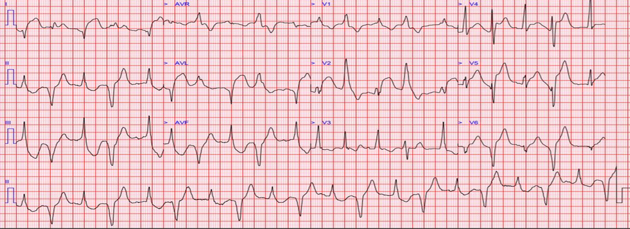
It's a typical ED shift. A nursing colleague passes you an ECG. The presenting complaint is chest pain. Of a set of borderline-looking strips, which would you activate PPCI for?

The answer is ALL of them. The exercise isn't a trick: it's demonstrating that hesitating on a borderline STEMI-equivalent costs tissue. "Time is tissue": the cost of over-calling PPCI is a discussion with cardiology; the cost of under-calling it is a bigger infarct.

Session syllabus for this topic: what the literature currently says about UK vs. US STEMI criteria (a young man with 2mm of ST elevation in V2+V3 can trigger the cath lab in the UK, but not in the US); the flaws in current STEMI definitions; the non-ECG factors that influence a PPCI consultant's decision when you call them; and what's actually going through their mind on that call.
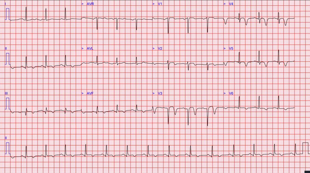
Anterior STEMI ECG
Anterior STEMI
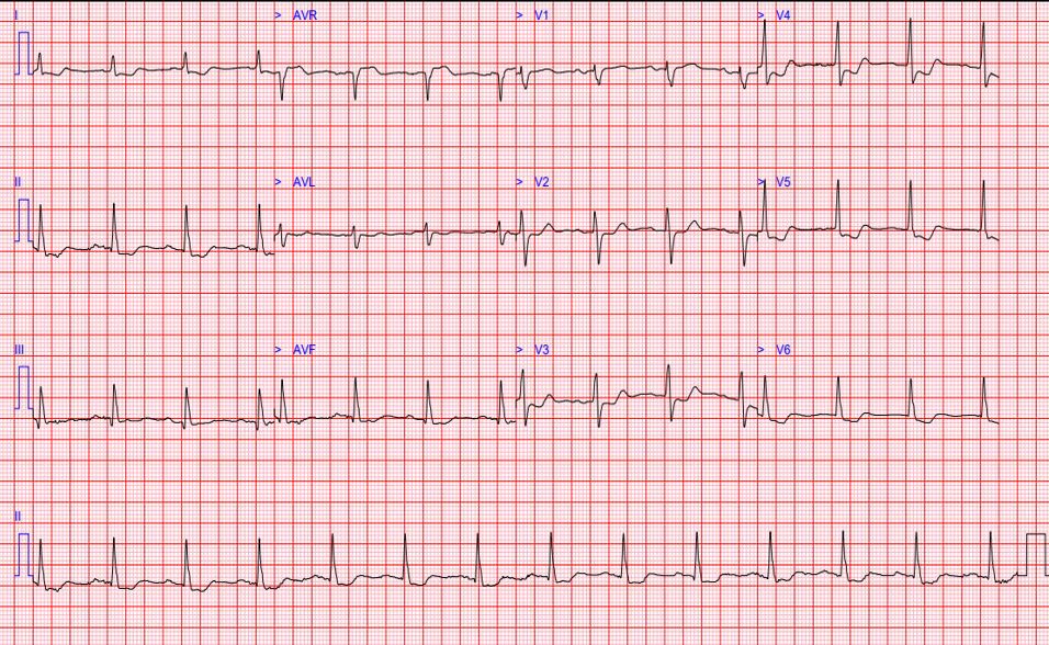
Inferior STEMI ECG
Inferior STEMI
Day 1 · Case

The hidden STEMI

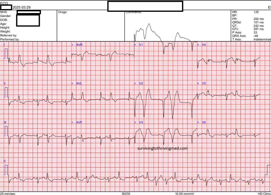
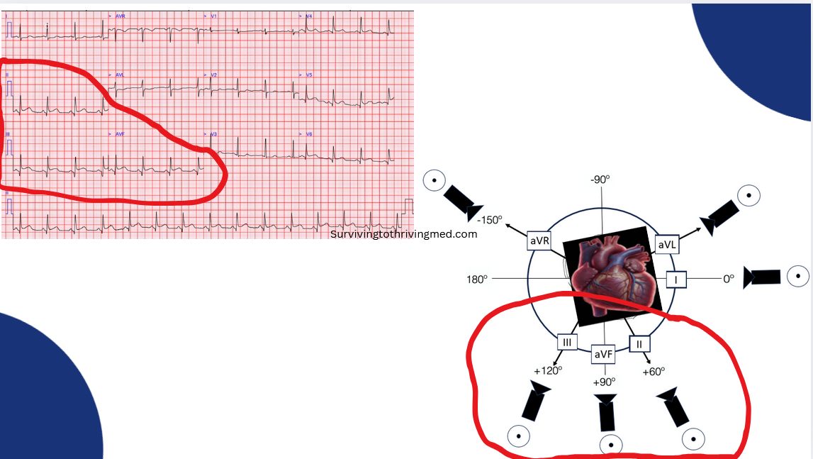
Sometimes the STEMI is genuinely present on the ECG, but goes unrecognised simply because the reader isn't aware of how the injury vectors work for that particular occlusion. This session covers the recognised STEMI-equivalents (patterns that meet PPCI criteria without classic, obvious ST elevation) and how to find them systematically rather than by pattern-matching from memory.

Wellens syndrome ECG
Wellens syndrome: a STEMI-equivalent hiding in the T waves, not the ST segment.
aVR ST elevation ECG
ST elevation in aVR: the pattern that points to left main or proximal LAD occlusion.
Day 2 · Case

VT, or not VT?

No arrhythmia workshop is complete without this question. The session works through VT vs. SVT-with-aberrancy from first principles (the pathophysiology and the literature behind the diagnostic tools available, and their caveats), so the reader can make an informed call rather than defaulting to a rule of thumb.
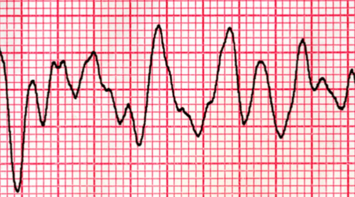
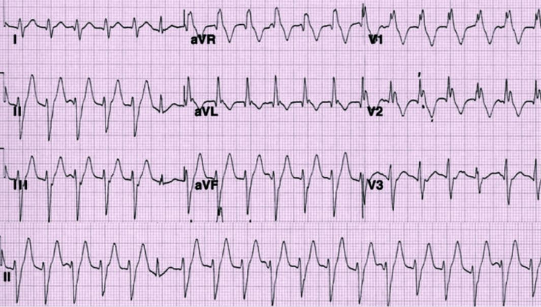
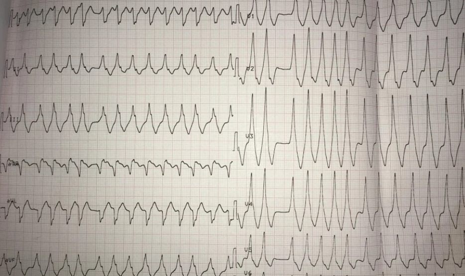
A set of broad-complex tachycardia strips is shown, each superficially resembling a "typical VT" pattern. The answer is NONE of them should get reflexive amiodarone. Amiodarone is not a universal cure for arrhythmias: there are situations (certain pre-excited AF, some polymorphic rhythms) where giving it blindly will make things worse, hard.
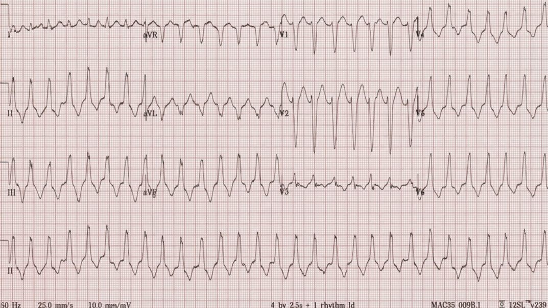
Monomorphic VT ECG
Monomorphic VT
Fascicular VT ECG
Fascicular VT
RVOT VT ECG
RVOT VT
Bidirectional VT ECG
Bidirectional VT
Pre-excited AF ECG
Pre-excited AF: irregular, varying morphology, and exactly the rhythm the callout above is warning about.
Day 2 · Case

Bundle branch blocks

Mnemonics like "William" and "Marrow" (for identifying LBBB and RBBB by QRS shape in V1/V6) are crutches that substitute for actually understanding the underlying physiology. Once you understand which bundle branch is blocked, and therefore which ventricle depolarises late and from where, the QRS morphology follows from that, rather than needing to be memorised as a shape to pattern-match.

See Part 8 — The Q wave for the septal depolarisation physiology this builds on.
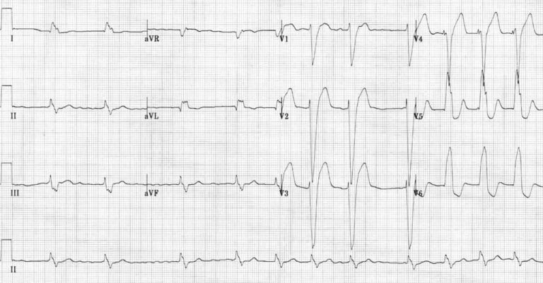
LBBB ECG
LBBB
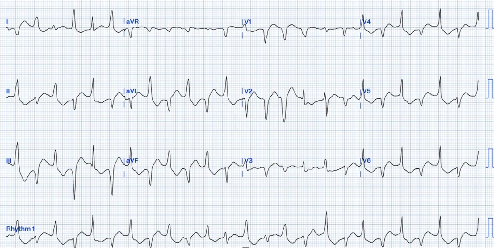
Bifascicular block ECG, nicknamed the South African flag sign
RBBB + LAFB: the "South African flag" pattern.
← ECG Fundamentals Back to Resources →

Already have an account? Log in →

No account yet? Sign up →